
To detect intracellular phosphorylated antigens by flow cytometry it is necessary to permeabilise and fix a cell population in a particular manner. This is necessary not only for phosphorylated proteins but any to maintain any surface immunophenotyping of cells.
Becton Dickinson have a human T-cell activation kit which allows the measurement of 6 phosphoproteins including, p38MAPK, Erk 1/2, Stat1, Stat3, Stat5 and Stat6. These antibodies are all directyl conjugated with Alexa Fluor 647. A pdf of the BD Phosflow T cell Activation Kit instructions are available from the link below.
The permeabilisation process makes use of 70% ice-cold methanol to allow antibody labeling of phosphoproteins. For surface labeling of lymphocyte subsets the T cell activation kit is supplied with a antibody cocktail of CD3-PerCP, CD4-FITC and CD8-PE. BD has also tested a range of their antibodies for surface labeling after methanol fixation. The pdf below gives a list of compatible surface labeling antibodies
Fluorophore compensations are set using the BD Compensation Bead technology with the control antibodies supplied in the kit. Instructions on there use are found on the link Compensation
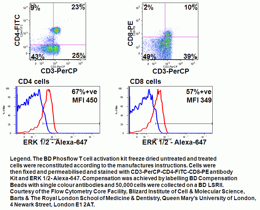
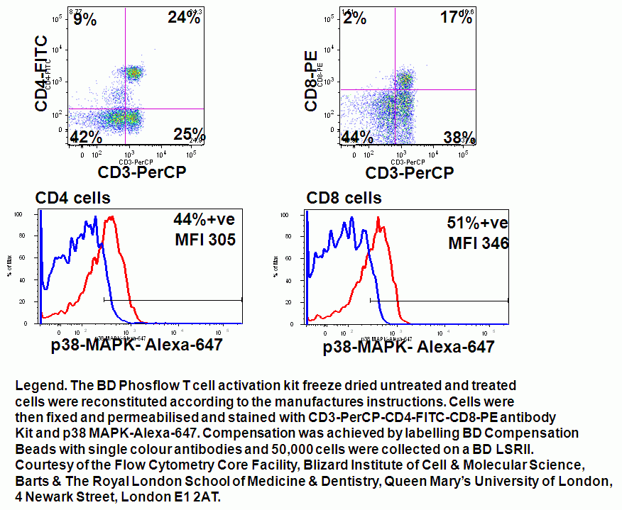
The treated and untreated control cells supplied in the T cell activation kit were separately stained for ERK 1/2 and p38 MAPK-Alexa-647 and the resulting histograms from CD4 and CD8 cells analysed by histogram overlays of the resting and treated cells.
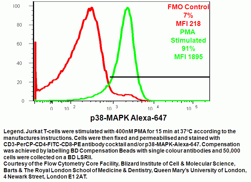
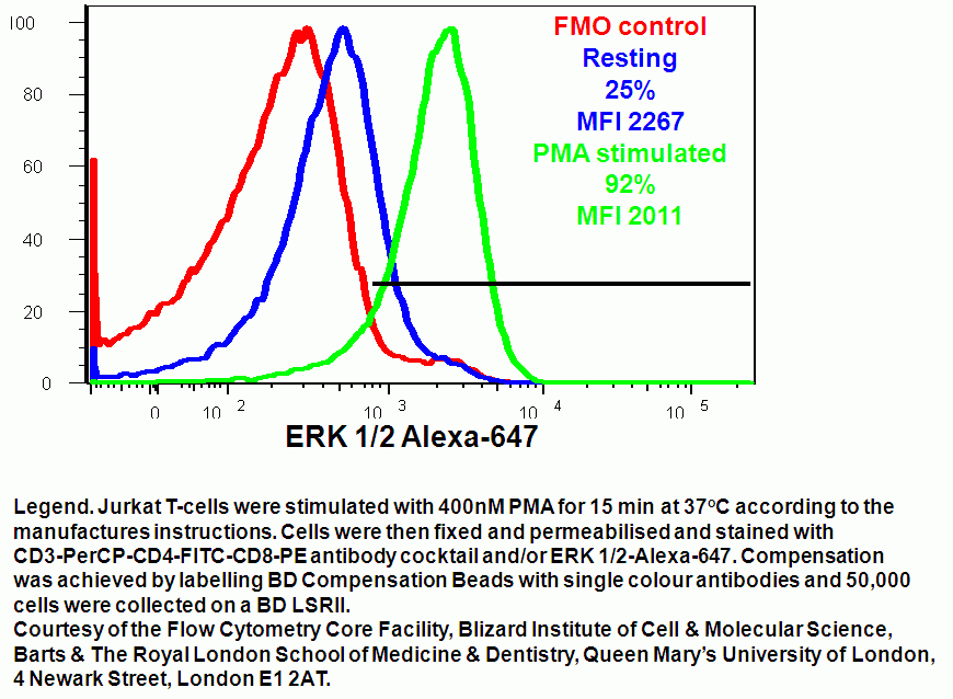
Jurkat T cells were stimulated for 15 minutes at 37C with 400 nM PMA and compared to unstimulated cells. Cells were then stained for CD3, CD4, CD5 and/or p38-MAPK-Alexa-647 or ERK 1/2-Alexa-647.
BD also have special software available on the internet called Cytobank which allows easy analyse of complex samples not only generating dot-plots, histogram overlays, tabulation of results but compares upregulation of phosphoproteins in terms of fold increase in expression and heat maps for easy display of data, see pdf.
